Accessory (OPA)
Optics Plate Accessory (OPA)
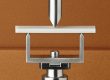
The OPA is an open optical system that permits basic visualization of sample structure during rheological experiments, revealing important insights about material behavior under flow. An open platform with a borosilicate glass plate provides a transparent optical path through which the sample can be viewed directly. This enhances the understanding of a range of materials, especially suspensions and emulsions. The accessory is easy to use and install, accommodates diverse optical systems, and offers accurate temperature control over a wide range for flow visualization and microscopy.
Features and Benefits
- Smart Swap technology for quick installation
- Simultaneous rheological measurements and direct visualization
- Visual access to any position within the measurement area, e.g. center, edge, or mid-radius
- Upper Heated Plate (UHP) with patented Active Temperature Control for precise temperature measurement
Technology
The OPA mounts to the DHR Smart SwapTM base and may be coupled with the Upper Heated Plate with Active Temperature Control for accurate, direct sample temperature measurement and control from -20 °C to 100 °C. The OPA can be used with cone or parallel plate geometries up to 60 mm in diameter.
The OPA is available in any of the following configurations:
- Open Plate: An open system that facilitates customization including a set of 8 M2 tapped holes for the easy adaptation of any optical system
- OPA with Modular Microscope Accessory (MMA): A static optical stage for microscopy.
- OPA with Digital Microscope: A high resolution digital camera permits the capture of still images or video. The camera is mounted on a y-z positioning stage to adjust focus and the field of view. Sample illumination is provided by the microscope’s 8 white LEDs.
Microscopy
OPA with Digital Microscope

OPA with Digital Microscope
The images below show the structure of a PDMS-PIB emulsion at rest and after shear flow. At rest, the emulsion structure consists of spherical droplets with a polydisperse size distribution. After shearing at 10 s-1 for 10 minutes, there is a decrease in the number of larger droplets and a shift towards more uniform droplet sizes.

MMA: Cross-Polarization Microscopy
MMA: Cross-Polarization Microscopy
The figures to the right shows an example of cross-polarization microscopy data collected using the Modular Microscope Accessory. A light crude oil sample was cooled from 25 °C to 0 °C at a controlled rate of 1 °C/min during a small amplitude oscillatory test. The images in the inset show cross-polarization micrographs of the sample at various stages during the experiment. At 20 °C, the sample is a homogeneous, low viscosity liquid with no crystalline features. As the sample is cooled, the viscosity rise sharply beginning at 15 °C. This process, known as outwaxing, is caused by the crystallization of long-chain hydrocarbons and paraffinic wax components in the sample and is accompanied by the appearance of several crystalline elements in the micrograph image. With additional cooling, the sample viscosity continues to increase, concurrent with an increase in the number and size of crystalline domains. Simultaneous imaging confirms that the cause of the observed viscosity increase is the onset of crystallization. The results highlight the use of microscopy as a powerful tool to investigate and understand the relationship between sample structure and its material properties.
MMA: Brightfield & Fluorescence Microscopy

MMA: Brightfield & Fluorescence Microscopy
The micrographs below demonstrate the imaging capability of the MMA in brightfield and fluorescence microscopy modes. The images show glass spheres suspended in polydimethylsiloxane (PDMS) in brightfield imaging, and fluorescently dyed polystyrene spheres dispersed in a commercial hair gel sample imaged using the dichroic splitter for fluorescence microscopy. A 20x objective was used to image both samples, scale bar = 50 μm.